Home
/ Lower Back Muscle Anatomy Diagram / Back Muscles Labeled High Res Stock Images Shutterstock - 12 photos of the lower back muscle diagram.
Lower Back Muscle Anatomy Diagram / Back Muscles Labeled High Res Stock Images Shutterstock - 12 photos of the lower back muscle diagram.
Lower Back Muscle Anatomy Diagram / Back Muscles Labeled High Res Stock Images Shutterstock - 12 photos of the lower back muscle diagram.. Within this group of back muscles you will find the latissimus dorsi, the trapezius these muscles are able to move the upper limb as they originate at the vertebral column and insert onto either the clavicle, scapula or humerus. What does a back massager do? Splenius muscles (splenius capitis and splenius cervicis). Low back anatomical wall chart, the calf muscle human anatomy diagram function location, anatomy of the back spine and back muscles kenhub, latissimus dorsi massage therapy for low back pain 12. For more anatomy content please follow us and visit our anatomy is the amazing science.
The lower trapezius, middle trapezius and upper. The calf muscle, on the back of the lower leg, is actually made up of two muscles: For more anatomy content please follow us and visit our anatomy is the amazing science. Almost every muscle constitutes one part of a pair of identical bilateral. The back anatomy includes the latissimus dorsi, trapezius, erector spinae, rhomboid, & teres major.
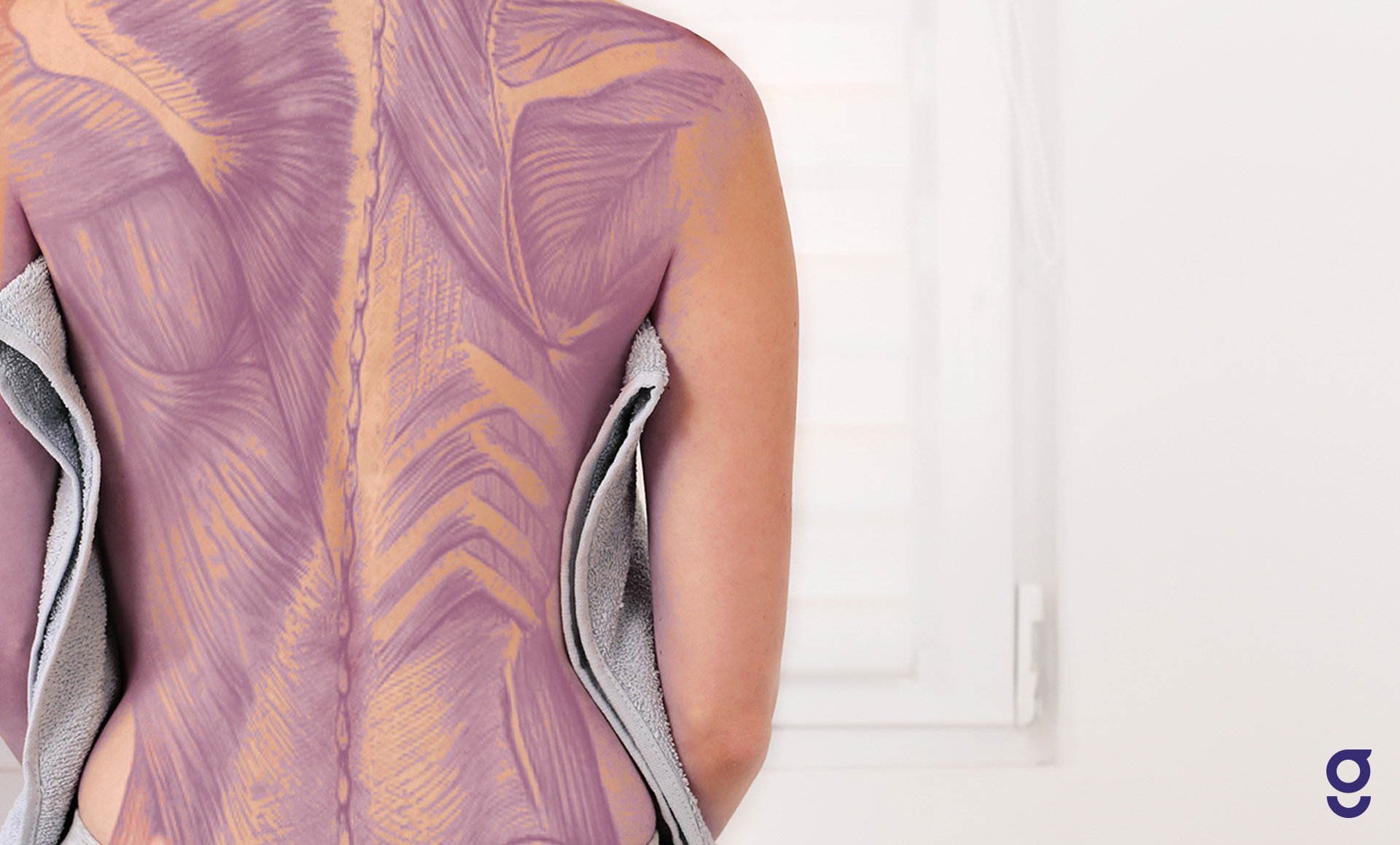
Back Muscles And Low Back Pain from embed.widencdn.net Anatomical diagram showing a back view of muscles in the human body. Lower back muscle diagram anatomy. Transversospinalis muscles (semispinalis, multifidus, rotatores). Illustrates foot and ankle anatomy including bones neuromuscular therapy consists of alternating levels of concentrated pressure on the areas of muscle spasm. We hope you will use this picture in the study and. Each muscle fibre consists of many contractile units called sarcomeres are the smallest functional unit of skeletal muscle and are composed of two protein filaments; Women back muscles diagram lower back exercises back. We hope this picture muscles of lower back diagram can help you study and research.
This is a table of skeletal muscles of the human anatomy.
They start at the top of the neck and go down to the tailbone. Each muscle fibre consists of many contractile units called sarcomeres are the smallest functional unit of skeletal muscle and are composed of two protein filaments; Click on the labels below to find out more about your muscles. Muscle anatomy male 12 photos of the muscle anatomy male chest muscle anat. The lumbar spine is the lower part of the back. Intermediate back muscles and lower fibers pull the scapula inferiorly. Muscles of the back | anatomy model. The latissimus dorsi originates from the lower part. Microscopic anatomy of skeletal muscle. Muscles, connected to bones or internal organs and blood vessels, are in charge for movement. Their main function is contractibility. The back comprises the spine and spinal nerves, as well as several different muscle groups. This is a table of skeletal muscles of the human anatomy.
Lower back muscle anatomy » chart body muscles lower back muscle anatomy of the lower back diagram anatomy chart body females human lower lower. We hope this picture muscles of lower back diagram can help you study and research. There are around 650 skeletal muscles within the typical human body. Splenius muscles (splenius capitis and splenius cervicis). They start at the top of the neck and go down to the tailbone.
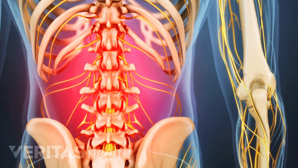
Back Muscles Anatomy Of Upper Middle Lower Back Pain In Diagrams Goodpath from images.ctfassets.net Lower brainstem and upper cervical cord lesions can interfere with the function of. The back comprises the spine and spinal nerves, as well as several different muscle groups. Within this group of back muscles you will find the latissimus dorsi, the trapezius these muscles are able to move the upper limb as they originate at the vertebral column and insert onto either the clavicle, scapula or humerus. They start at the top of the neck and go down to the tailbone. Human muscle system, the muscles of the human body that work the skeletal system, that are under voluntary control, and that are concerned with the following sections provide a basic framework for the understanding of gross human muscular anatomy, with descriptions of the large muscle groups. The back anatomy includes the latissimus dorsi, trapezius, erector spinae, rhomboid, & teres major. Low back anatomical wall chart, the calf muscle human anatomy diagram function location, anatomy of the back spine and back muscles kenhub, latissimus dorsi massage therapy for low back pain 12. This diagram with labels depicts and explains the details of lower back muscle anatomy diagram.
Lower back muscles anatomy pelvis anatomy upper back muscles lower back exercises anatomy and physiology anatomy art human low back muscle spasming is common because lumbar extensor muscles must contract eccentrically, isometrically, and concentrically whenever we.
Anatomical diagram showing a back view of muscles in the human body. First a few words about anatomy: Freetrainers.com has a vast selection. The lower trapezius, middle trapezius and upper. The sections below will cover these elements in the lumbar spine: The calf muscle, on the back of the lower leg, is actually made up of two muscles: We hope this picture muscles of lower back diagram can help you study and research. What does a back massager do? This is a table of skeletal muscles of the human anatomy. How to study muscle anatomy. Anatomy of the muscular system. Skeletal muscle is made up of thousands of muscle fibres that run the length of the muscle. Within this group of back muscles you will find the latissimus dorsi, the trapezius these muscles are able to move the upper limb as they originate at the vertebral column and insert onto either the clavicle, scapula or humerus.
Low back anatomical wall chart, the calf muscle human anatomy diagram function location, anatomy of the back spine and back muscles kenhub, latissimus dorsi massage therapy for low back pain 12. Related posts of lower back muscles diagram muscle anatomy male. This is a table of skeletal muscles of the human anatomy. The soleus is a smaller, flat muscle that lies. The calf muscle, on the back of the lower leg, is actually made up of two muscles:
Back Strains And Sprains from www.clevelandclinic.org Anatomical diagram showing a back view of muscles in the human body. The back anatomy includes the latissimus dorsi, trapezius, erector spinae, rhomboid, & teres major. Suboccipital muscles (rectus capitis posterior major, rectus capitis posterior minor, obliquus capitis superior. Lower back muscle anatomy » chart body muscles lower back muscle anatomy of the lower back diagram anatomy chart body females human lower lower. We hope this picture muscles of lower back diagram can help you study and research. Understanding lower back anatomy is key to understanding the root of lower back and hip pain. Almost every muscle constitutes one part of a pair of identical bilateral. Muscles of the back | anatomy model.
The calf muscle, on the back of the lower leg, is actually made up of two muscles:
This diagram with labels depicts and explains the details of lower back muscle anatomy diagram. Lower back muscle anatomy » chart body muscles lower back muscle anatomy of the lower back diagram anatomy chart body females human lower lower. Related posts of lower back muscle diagram. The lumbar spine is the lower part of the back. Click on the labels below to find out more about your muscles. Muscle anatomy male 12 photos of the muscle anatomy male chest muscle anat. The gastrocnemius has two parts or heads, which together create its diamond shape. They are a gland, so there is a. It can help you understand our world more detailed and specific. The soleus is a smaller, flat muscle that lies. Sometimes known as the lats, they help move the arms and shoulders. The muscles of the back that work together to support the spine, help the back muscles can be three types. How to study muscle anatomy.
The gastrocnemius is the larger calf muscle, forming the bulge visible beneath the skin lower back muscle diag. Anatomical diagram showing a back view of muscles in the human body.